There are many imaging modalities available to health professionals which can assist with the diagnosis and management of injury or pathology.
Types of imaging may include MRI scans, x-rays and CT scans, and the use of these modalities is often at the discretion of your healthcare professional, whether that be a physiotherapist or doctor.
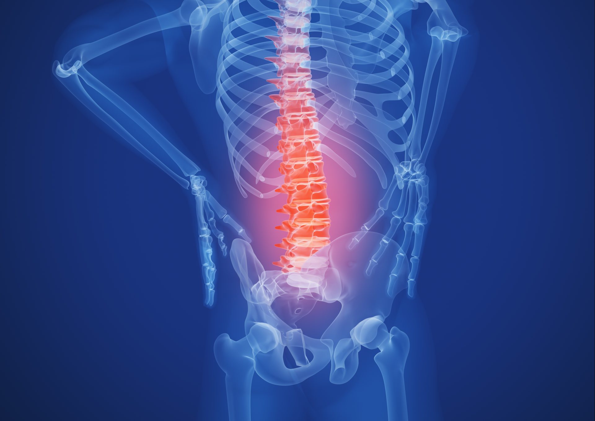
When it comes to MRI reporting, a large proportion of the population who have no pain also have findings considered abnormal.
In a review of over 3000 people who had a lower back MRI performed, a majority had findings which would be considered atypical1.
The interesting part of this study is all participants had no reported pain or disability. These results are outlined in the table below.
Lower back MRI in asymptomatic individuals
| Age | Disc degeneration | Disc bulge | Disc protrusion |
| 20 | 37% | 30% | 29% |
| 30 | 52% | 40% | 31% |
| 40 | 68% | 50% | 33% |
| 50 | 80% | 60% | 36% |
| 60 | 88% | 69% | 38% |
| 70 | 93% | 77% | 40% |
| 80 | 96% | 84% | 43% |
As you can see, by the age of 40 most people are showing disc ‘degeneration’ and bulges, even when they have not reported any pain.
Looking at these numbers it may mean we need to reconsider what is considered normal particularly as we move through our life stages.
These findings are not limited to the lower back either. A similar study was done on the knees of asymptomatic individuals in 2018.
The MRI performed on over 4,000 people suggested that 43% of people over 40 had cartilage defects and osteoarthritis (OA) despite no pain2.
What these studies outline is the importance of knowing what an imaging report tells us.
It’s important to remember that while an MRI may report findings on your area of pain, a large proportion of these findings are normal and found in people without pain.
‘Atypical’ scans are often not something to worry about in isolation, the real challenge for a physio like myself, is to work out why the area has become painful and what the best approach may be to manage and reduce the pain.
References
- Brinjikji et al. Systematic literature review of imaging features of spinal degeneration in asymptomatic populations. Am J Neuroradiol. 2015 Apr;36(4):811-6.
- Culvenor et al.Prevalence of knee osteoarthritis features on magnetic resonance imaging in asymptomatic uninjured adults: a systematic review and meta-analysis. British Journal of Sports MedicinePublished Online First:09 June 2018. doi: 10.1136/bjsports-2018-099257