Low back pain is one of the most common causes for disability in Australia and it’s important to note that its presentation and severity can vary enormously.
At Lifecare Prahran Sports Medicine a common question our sport and exercise medicine physicians and physiotherapists must answer when working with patients presenting with low back pain is, ‘Do I need a scan?’
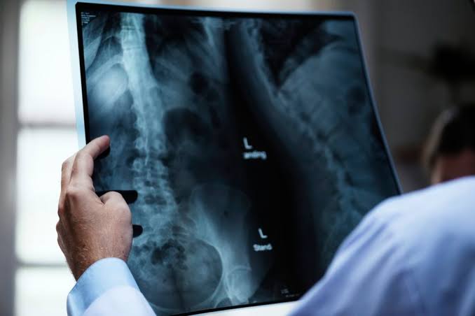
Whilst to most people, an x-ray or MRI might seem a useful or logical aid in formulating a diagnosis, in a significant percentage of cases scans are neither required nor indicated, and can at times actually create more fear and in fact slow patient recovery!
In a recent published article (BJSM 2019 – O’Sullivan et al – see below), scans of the lumbar spine were found to be associated with greater levels of reported pain, disability, work absenteeism, medication use and healthcare seeking.
When assessing patients with insidious onset (no specific trauma) of lower back pain, imaging such as x-ray or MRI is typically only indicated where a patient presents with signs suggestive of a more serious cause for their pain such as infection, stress fracture, malignancy or significant nerve compression (BMJ 2001 – Kendrick et al).
Certain features within the history and physical examination performed by your treating practitioner will attempt to screen for such conditions.
If it is felt that imaging is required or beneficial, it then comes down to choosing the modality most appropriate for the presentation.
This all sounds easy and many patients (and even lesser experienced health practitioners) believe MRI to be the ‘best investigation for everything’.
In reality, there are often cases where other modalities such as x-ray, CT (CAT) scan, nuclear bone scan (and occasionally even ultrasound), can be far more useful.
Certain medical conditions (e.g. pregnancy, cardiac pacemaker) might preclude some modalities but not others.
It’s important to understand some of the issues surrounding the use of imaging in the investigation of lower back pain, and in turn appreciate why imaging is not always a beneficial thing.
Imaging modalities such as x-ray/CT and bone scan involve the use of ionising radiation and thus the potential benefits of information obtained from the investigation(s) must be weighed up against any potentially negative health effects from radiation exposure.
This is even more relevant in the case of a pregnant or potentially pregnant female.
Depending on the imaging facility and/or the conditions being investigated, imaging investigations can be expensive.
Some scans involve injections of contrast and this intervention can also pose theoretical risk of allergic reactions or complications from the injection itself.
A concept increasingly on the rise in terms of awareness, and of equal importance to the issues discussed above, is that of age-relevant incidental findings.
Much the same as a car might show signs of rust or loss of springiness in the seats as it ages, the musculoskeletal system naturally wears out with use too.
This can result in degenerative changes throughout any region, and as one would expect, the likelihood of finding these changes on scans increases with age.
The fact that numerous studies have shown the incidence of degenerative changes throughout the musculoskeletal system (and especially the lumbar spine) to be high in the asymptomatic population (Brinjikji et al 2015 is a good reference- see below) further adds to the conundrum that health practitioners face in terms of interpreting scan findings.
It is actually expected that most patients over the age of 30 will have ‘changes’ on their scans of the lumbar spine.
The problem arises where attempts are made to make the diagnosis ‘fit’ these changes.
Such scan findings could in fact often be considered false positives; not the cause of the patients symptoms or pain – a so-called ‘red herring’.
These changes certainly don’t predict pain.
Difficulties can also arise where scans reported as ‘abnormal’ lead to additional tests or investigations being ordered, in turn adding to the stress of the patient by means of further radiation, cost, visits to the health practitioner and the overall uncertainty and fear surrounding their diagnosis.
Of course sometimes these additional tests are indicated and this is where the expertise of our sport and exercise medicine physicians and physiotherapists makes all the difference.
Once your treating practitioner is satisfied they have excluded serious spinal pathology, the rest of their assessment and treatment aims to target the mechanical or inflammatory nature of your pain.
It is important to note that for the majority of non-specific changes seen on spinal scans, the mainstay of treatment is conceptually the same – promotion of activity and mobility, avoiding dependence on analgesic medications and continued work on improving strength and endurance through the spine and its supports.
This is yet another reason why, for the majority of presentations of lower back pain scans, are not actually required!
Our highly experienced sport and exercise medicine physicians and physiotherapists will help you get the fastest, most comfortable recovery possible and assist in improving both your general quality of life and a prompt and safe return to work, whilst also targeting a graduated return to your chosen sports and recreations as soon as possible.
Most importantly however, our team are here to listen to you, and will always have time to address any concerns you may have regarding the appropriateness of imaging or any other feature of the overall management of your back pain.
Working together with you they will help formulate a mutually agreeable plan and help you get the diagnosis, reassurance and recovery you’re after.
Three useful links from this blog:
- O’Sullivan K, O’Sullivan PB, O’Keeffe M. The Lancet series on low back pain: reflections and clinical implications.
- Kendrick D, Fielding K, Bentley E et al (2001) Radiography of the lumbar spine in primary care patients with low back pain: randomised controlled trial. BMJ 322:400–405. https://doi.org/10.1136/bmj.322.7283.400
- Brinjikji W, Luetmer PH, Comstock B, et al. Systematic literature review of imaging features of spinal degeneration in asymptomatic populations. AJNR Am J Neuroradiol 2015;36(4):811–16. doi: 10.3174/ajnr.A4173. https://www.ncbi.nlm.nih.gov/pubmed/25430861
Dr Stuart Down is a Sport & Exercise Medicine Physician. He works at Lifecare Prahran Sports Medicine on Monday, Wednesday and Friday.
Eric Coleman is an APA Titled Musculoskeletal Physiotherapist. He works at Lifecare Prahran Sports Medicine on Tuesday, Wednesday, Friday and Saturday.
The clinic is close to suburbs including Malvern, South Yarra, Toorak, Armadale, St Kilda East, Caulfield, Richmond and Hawthorn, and has early and late appointments for all your sports medicine and physiotherapy needs.