Bone is a dynamic tissue that is constantly remodelling under the influence of multiple mechanical and hormonal influences.
Under normal circumstances in healthy conditions, bone resorption (bone ‘removal’ – performed by cells called osteoclasts) is in equilibrium with bone synthesis and formation (carried out by other cells known as osteoblasts).
It is normal for minor micro-fractures within bone to self-repair when adequate rest is provided or where loads on the bone are continually sub-threshold.
In these cases, more bone is often laid down as reinforcement in areas of stress (this can be seen on various imaging scans as ‘sclerosis’ of bone).
If however, a bone is subjected to continual, repetitive loads or increases in load above and beyond that which normal bone resynthesis can cater for, bone damage ensues.
Accumulation of bone marrow oedema occurs in increasing intensity through the periosteum and /or marrow cavity (referred to as bone stress / stress reaction and not stress fracture (as there is no cortical breach) (1), though micro-fractures can propagate, culminating in the formation of stress fractures.
Furthermore, there is typically a lag between the resorptive process and the subsequent influx of bone-laying osteoblasts, and when coupled with continual high intensity loading and in particular rapid increases in load, bones are particularly vulnerable to stress fracture formation.
Generally speaking, there are two categories of stress fractures – fatigue fractures and insufficiency fractures.
Fatigue fractures arise from excessive loading of bones with normal mineralisation.
This group is characteristically (but not exclusively) seen in the younger age group (under 50) who are engaging in repetitive high loading activity.
Insufficiency fractures on the other hand, arise when those with abnormal, osteoporotic bone are subjected to what is generally considered an acceptable amount of load.
This latter group is typically seen in older patients, more so females, or patients with chronic medical conditions or long term use of cortisone medication.
Another scenario where insufficiency stress fractures can be seen is where bones are not subject to enough load and this can result in relative ‘disuse osteopenia’ (impaired bone density).
This might be from prolonged immobility (e.g. lengthy periods of confinement to bed from ill health / prolonged use of crutches / people who are wheelchair bound or with spinal cord injuries and even in astronauts who are in space for lengthy periods without gravitational force and weight bearing).
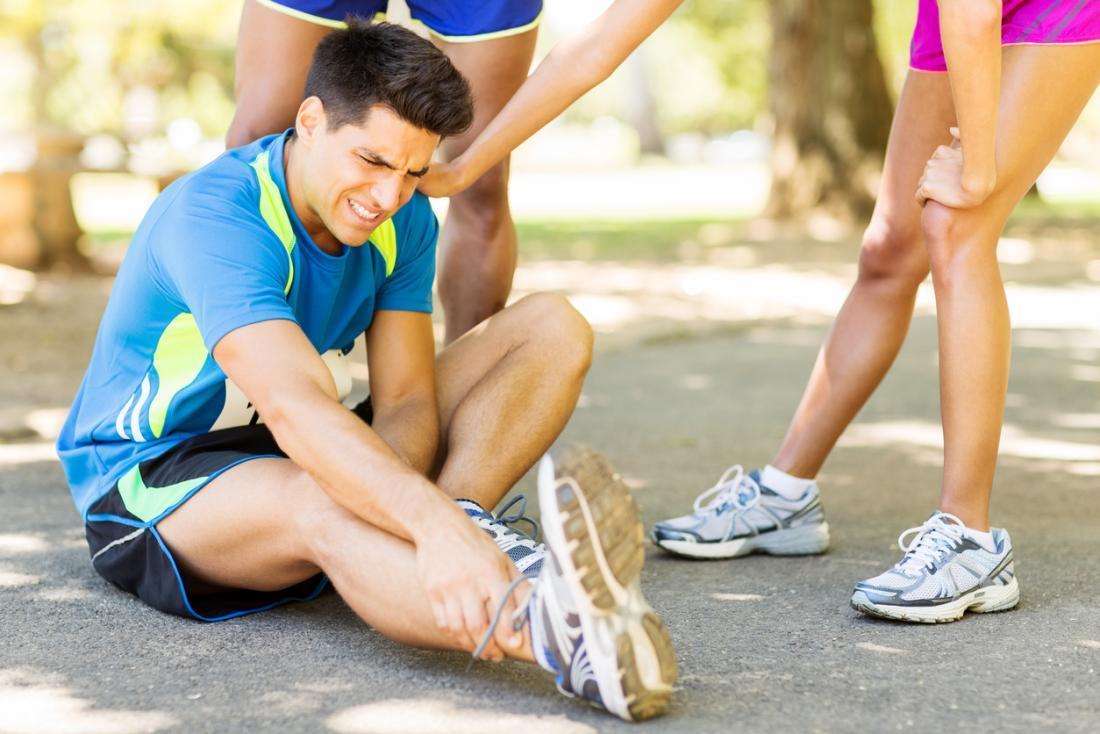
Stress fractures are particularly common in the bones of the foot, leg, and pelvis.
These bones are required to absorb the forces and impact created from walking, running, and jumping.
Up to 12 times the weight of the body may be generated with each step, and our bones, joints, muscles, and ligaments work together to cushion the body against that force.
This highlights the importance of a sound ‘kinetic chain’, whereby each link in the chain of movement are optimal and as such, helping one another absorb the forces.
If one or more links falter, the remaining links have to carry the burden, and may fail.
Stress fractures of the upper limb and chest are much less common (2) and are typically a lot more sport-specific in presentation.
Humeral bones stress in tennis players and baseball pitchers, rib stress fractures in rowers and coracoid process stress fractures in trap-shooters (3) are well documented examples (3, 4).
Stress fractures can be further categorised in clinical practise, into low risk and high risk variants.
Low risk fractures are those which typically heal well and promptly with appropriate rest.
The high risk group however, are typically found in anatomic sites with a predilection for slow or incomplete healing, a high tendency for recurrence, or a significant risk of complication with fracture progression (5).
This group is also seen more so on the tension side of long bones (e.g. superior femoral neck / anterior tibial cortex).
X-rays may often appear normal, especially in the earlier stages of bone stress, where there may only be bone oedema (bruising) present, but not yet cortical disruption or periosteal reaction (new bone formation).
X-ray abnormalities can usually be best appreciated beyond 3-4 weeks of symptomatic bone stress. Generally, MRI is considered the best means of detecting bone stress injury / stress fractures, and is the investigation of choice for many stress fractures, especially those stress fracture subtypes considered high risk.
MRI has the advantage of stratifying severity of bone stress, confirming the presence or absence of an associated fracture line (in turn providing better estimates for expected return to weight-bearing and sporting activity), and can also be useful in diagnosing other conditions that may present as bone stress injury (e.g. joint synovitis / bone tumours / avascular necrosis).
MRI has the added advantage of no ionising radiation.
The cornerstone of management for all stress fractures is an adequate rest period to allow bone healing and reinforcement at the fracture site, a progressively graduated return to impact or the initially insulting activity along with attendance to any predisposing or precipitating issues that can often be clearly elicited in a carefully obtained history.
There will in most cases be a sudden increase in either or both of volume or intensity of activity, a change in running shoes or terrain (in the case of lower limb or pelvic stress fractures), or a preceding injury or region of stiffness that contributed to overload of the part in question
Sporting technique may need to be modified (e.g. changing in bowling delivery position for cricketers with lumbar stress fractures). All of these factors must be borne in mind for prevention of recurrences.
Attendance to biomechanical issues within the kinetic chain for that movement or action is also imperative.
For those with lower limb stress fractures, formal running assessment on a treadmill can be of great benefit and subtle changes to running technique can result in significant running efficiency gains and improvements of force dissipation with foot strike.
Where subjects have stress fractures of the lower limb and are unable to walk without pain, periods of ambulatory rest in a CAM boot for 2-6 weeks can be highly beneficial, whilst still allowing removal for sleep, personal hygiene and non impact cross training.
Generally there is no role for anti-inflammatory medications and some reports even suggest they may slow bone healing.
Standard analgesics (e.g. Panadol/Panadeine) early on, coupled with rest are usually more than sufficient to control pain.
In most cases, your practitioner will recommend continued pain-free non-impact cross training to maintain general fitness and muscle tone, though in some cases of higher risk, patients may be advise strict rest.
Management of stress fractures of the chest / back or upper limb usually requires no major changes to daily life, but marked reduction or even abstinence from the activity that led to the stress fracture.
Again, cross training with pain free activity is encouraged, and attention to biomechanical issues and training loads is imperative.
Some studies suggest low intensity pulsed ultrasound (LIPUS) to be beneficial in stress fracture healing (4, 5), whilst others do not support these findings.
Younger athletes with recurrent stress fractures (and especially females where menstrual and hormonal status can have effects on bone density) should consider the possibility of energy deficiency states (known as RED-S) leading to impaired bone density and consultation with a dietician is always prudent to assess caloric intake and diet in general.
In recurrent multi-regional stress fractures or patients of older age (>40), consideration should be given to investigation of bone density and management of any derangements found.
Dr Stuart Down is a Specialist Sport & Exercise Medicine Physician and has over 15 years of experience dealing with stress fractures and all manner of other activity-related injuries and conditions. To see Dr Down or any of his colleagues for assessment of exercise related shin or calf pain, phone Lifecare Prahran Sports Medicine on (03) 9529 8899.
Sources
- Fredericson M, Bergman AG, Hoffman KL, Dillingham MS. Tibial stress reaction in runners. Correlation of clinical symptoms and scintigraphy with a new magnetic resonance imaging grading system. Am J Sports Med 1995; 23:472-481.
- Matheson GO, Clement DB, McKenzie DC, et al. Stress fractures in athletes: a study of 320 cases. Am J Sports Med 1987;15:46–58.
- Boyer DW Jr, Trapshooter’s shoulder: stress fracture of the coracoid process. Case report. J Bone Joint Surg Am.1975 Sep;57(6):862.
- Grant Lloyd Jones, MD, Upper Extremity Stress Fractures, Clin Sports Med 25 (2006) 159-174 25 (2Sports Med 25 (2006) 159–174
- Boden BP, Osbahr DC. High-risk stress fractures: evaluation and treatment. Am Acad Orthop Surg 2000;8(6):344–53.