Throughout my physiotherapy studies, we were always told to trust our clinical assessment and judgement over medical imaging.
What shows up on a scan quite often correlates poorly to our patients clinical presentation and the radiographer’s report should only add to our findings, not supersede all others.
This is particularly true in low back pain and neck pain.
Recently a patient and close friend of mine Thomas Brennan, sustained a ‘significant’ soft tissue injury.
His report did not read well, but as he bounced through the waiting room into my office, it was clear he was doing better than the scan suggested.
He later proved this recommendation true, that we should trust our clinical judgement before making a decision. I thought I’d share his success as a case study.
History
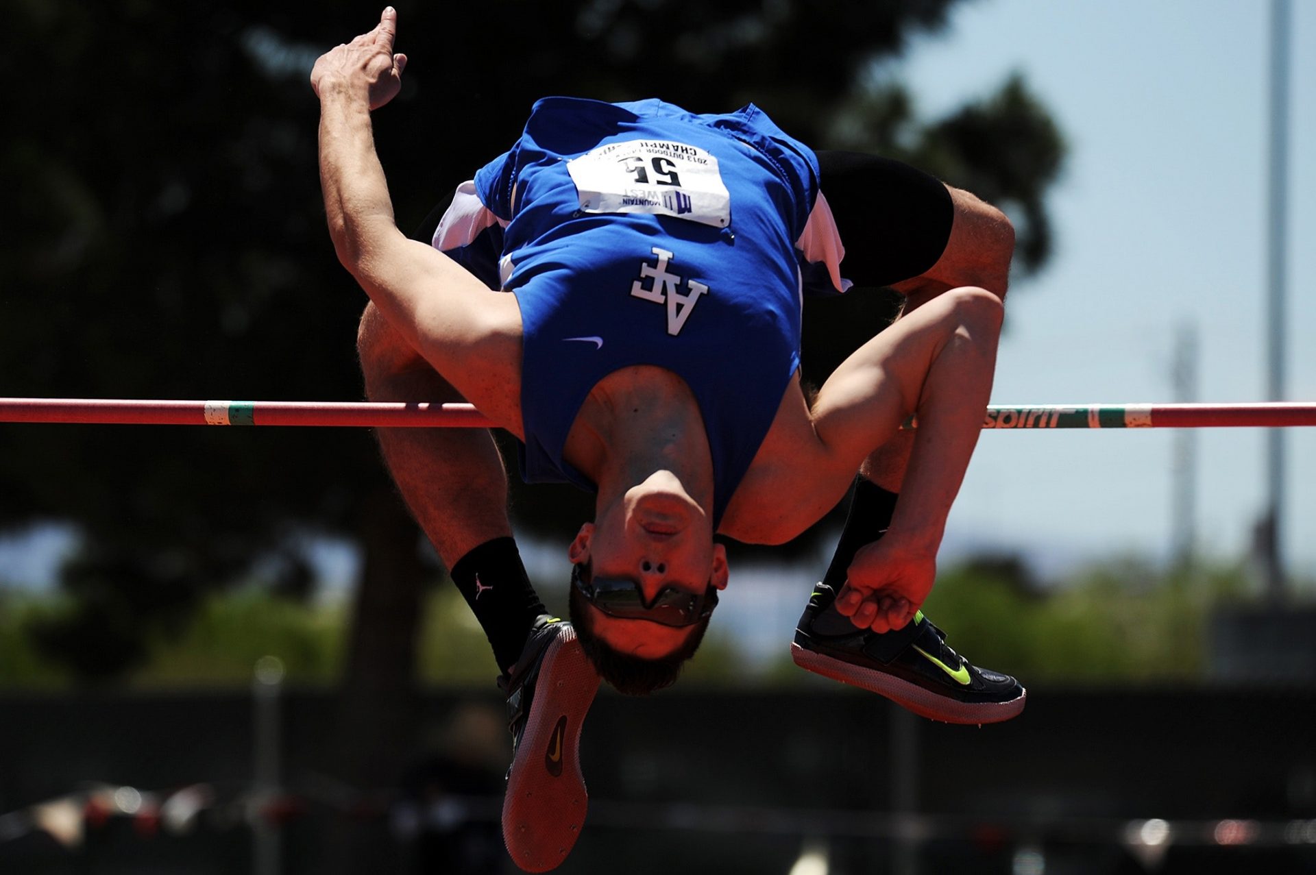
I received a call from Tom over the weekend while he was competing in a high jump meet over east.
He explained that he felt a pull in his left calf when performing a scissor kick.
He immediately knew he’d sustained an injury, but wasn’t too concerned thinking it was hopefully just a grade 1 strain.
The priority for Tom was to be fit for Nationals in approximately 5 weeks, so we sent him for an MRI immediately.
Tom’s MRI report read: Extensive (BAMI Grade 3c) proximal tendinous tear of the lateral soleus.
At first glance, this didn’t read well at all, reporting an extensive muscle tear affecting greater than 50% of the muscle and involving the soleus tendon (3.5cm of the tendon to be exact).
Considering this was Tom’s jumping leg, my initial thought was that Nationals was likely going to be ruled out.
3 days after the injury, Tom walked into the clinic bouncing on his toes.
The same way he has done for as long as I’ve known him.
He had minimal pain, none when walking at a comfortable pace and he’d done all the early management he was advised (ice, rest, compression, active ROM etc.).
Tom didn’t look like someone who’d sustained a grade 3 tear (or at least what I was expecting to see).
Objective examination
- Tenderness on palpation lateral soleus (1 x 5cm)
- Calf raise (knee in full extension) – 15+ reps with mild discomfort
- Calf raise (knee in flexion) – 6 reps with moderate pain
- Knee to wall test – 7cm pain limiting
- Negative neural provocation tests (straight leg raise)
From the way Tom presented subjectively and objectively, we agreed this was something we would treat and plan for based on his clinical findings.
Although the MRI report suggested otherwise, we were both happy to monitor his progress before making a firm decision.
Ideally we wanted at least 2 weeks of full training and competition, prior to competing at Nationals.
Tom and I sought the opinion of Tom’s former athletics physio who agreed that we should progress based on his clinical findings, not our expectations based on the MRI.
Tom’s rehabilitation involved:
- Calf stretching
- Isometric calf raises with progression to eccentric calf exercise (determined by Tom’s symptoms and soreness following exercise)
- Closed kinetic chain exercises – squats, lunges
- Further progression to full range single leg calf raises on the edge of a step, single leg squats, eccentric only exercises targeting soleus and hopping exercises
Tom presented 10 days later to the clinic reporting, no pain with any exercise or daily activities.
Objectively he could hop, calf raise (gastrocnemius), calf raise (soleus) and single leg squat all without any symptoms.
At this point Tom and I liaised with his athletic coach and progressed through a plyometric program, running program and return to jumping program as tolerated (very well!).
Outcome
Tom managed to successfully return to competition in just over 3 weeks post injury.
Unfortunately, he didn’t jump as well as he would have liked at Nationals, as he was hindered by another previous injury.
However, he has not had any complications with his calf since the initial injury.
For me, this was a great success to see a well executed rehabilitation program and plan succeed beyond our initial expectations.
It was also a strong reminder of what I’d always been taught – medical imaging may add to the clinical picture, but in the end we should always trust our clinical judgement.
It could be the difference between an athlete competing in something they’ve been training for all year or missing it completely.
The difference between a premiership or sitting on the side lines.